Research Databases Serous Images
Microscopical Image Database (Serous cytology)
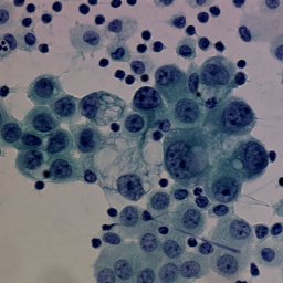
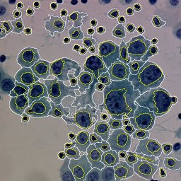
This database contains 10 color microscopic images from serous cytology.
A ground truth is also available for each image. Each region of the ground
truth region corresponds to a given nucleus and has a specific label.
The background has the label one. Figures below illustrate the purpose
of the image processing process: the extraction of cellular components
(left: original image, center: ground truth, right: segmented image).
Please Note that in this segmentation process, cytoplasm is also extracted
but is not available in the ground truth.

You can access to this database by the following link. Download the Serous Image Database.
You are free to use the dataset for non-commercial research and educational purposes. In exchange, we request only that you cite our IEEE trans. IP paper:
@article{Lezoray-2002,
author={O. Lezoray and H. Cardot},
title={Cooperation of color pixel classification schemes and color watershed :
a study for microscopical images},
journal={IEEE transactions on Image Processing},
volume={11},
year={2002},
pages={783-789},
number={7}
}
author={O. Lezoray and H. Cardot},
title={Cooperation of color pixel classification schemes and color watershed :
a study for microscopical images},
journal={IEEE transactions on Image Processing},
volume={11},
year={2002},
pages={783-789},
number={7}
}